
Can Liposuction Remove Stubborn Fat Even If You Are Not Overweight?

Can Liposuction Remove Stubborn Fat Even If You Are Not Overweight? A person’s weight does not always tell the full story of their body shape. Two individuals may have nearly identical measurements on the scale yet look noticeably different because of where their bodies store fat. Even people with a lean appearance may notice isolated areas of fullness that remain unchanged despite regular exercise and careful eating habits. For some, the concern is a small accumulation of fat below the belly button. Others may notice that their waist lacks definition or that their thighs appear fuller than the rest of their body. These differences are often related to inherited fat distribution patterns rather than an inability to maintain a healthy weight. Liposuction can remove stubborn fat even in people who are not overweight. The procedure is designed to selectively reduce subcutaneous fat deposits, making it possible to reshape particular body regions without pursuing substantial weight loss. However, successful treatment depends on more than identifying an unwanted fat deposit. Skin elasticity, fat thickness, underlying anatomy, and realistic expectations all influence whether liposuction is an appropriate option. Why Can Stubborn Fat Remain Even When You Are Slim? The human body does not distribute fatty tissue according to a person’s aesthetic preferences. Fat storage follows biological patterns influenced by genetics, hormones, and individual anatomy. This explains why some people develop fullness around the hips while others accumulate more fat along the lower abdomen or waist, even when their overall weight remains within a healthy range. Genetic factors influence both the location of fat deposits and the way different body regions respond to weight changes. Someone may lose weight from their face, arms, or upper body while retaining a relatively larger amount of fat around the thighs or flanks. These differences can become especially frustrating for individuals who already maintain a healthy lifestyle. Exercise improves cardiovascular health, supports muscle development, and contributes to overall fat reduction. However, the body does not selectively burn fat from whichever area is being exercised. For example, strengthening abdominal muscles does not guarantee that the fatty tissue covering them will disappear. Localized fat cannot be reliably eliminated through targeted exercises alone. Although total body fat may decrease with consistent physical activity and an appropriate diet, certain regions can remain proportionally fuller than others. Hormonal influences contribute to these patterns as well. Estrogen plays a role in fat storage around the hips and thighs, while changes associated with aging and hormonal balance can influence abdominal fat distribution. As people get older, changes in body composition may also make certain fat deposits more noticeable, even without significant weight gain. Another important consideration is that pursuing additional weight loss does not always improve body proportions. A person who is already slim may lose volume from areas they would prefer to maintain while seeing relatively little change in the region that concerns them. Stubborn fat in a person with normal body weight is not necessarily a sign of excess body fat. It may simply reflect the body’s natural pattern of fat distribution. For individuals who want to address these localized differences, surgical body contouring may offer an alternative to attempting further overall weight reduction. How Does Liposuction Change Body Shape Without Significant Weight Loss? The defining feature of liposuction is its ability to reduce fat in selected anatomical regions rather than attempting to change the body’s overall weight. During the procedure, a surgeon removes a controlled amount of fatty tissue from beneath the skin using a narrow instrument known as a cannula. The cannula is introduced through small incisions and used to extract fat from the planned treatment area. Unlike diet-related weight loss, which primarily reduces the volume of existing fat cells, liposuction physically removes a portion of the fat cells from the treated region. The resulting change can improve the relationship between neighboring body structures. For example, a person may have relatively narrow hips but a less defined waist because of localized fat along the flanks. Reducing this tissue may create a more noticeable waistline without substantially affecting body weight. Similarly, removing a modest amount of fat from beneath the chin may improve the transition between the jaw and neck. In such cases, the amount of tissue removed may be relatively small, yet the change in contour can be visually meaningful. The purpose of liposuction is not maximum fat extraction. It is the selective reshaping of body contours while preserving natural anatomical transitions. Several surgical methods may be used to accomplish this. Traditional suction-assisted liposuction removes fat through controlled cannula movement and suction. Power-assisted techniques use a mechanically vibrating cannula, while ultrasound-assisted approaches use ultrasonic energy to help loosen fatty tissue before removal. The choice of method depends on factors such as the treatment area, tissue characteristics, previous surgery, and the surgeon’s assessment. It is also important to understand that liposuction treats only certain types of fat. Subcutaneous fat, located immediately beneath the skin, can be removed through liposuction. However, visceral fat, which surrounds internal organs inside the abdominal cavity, cannot be extracted using conventional liposuction techniques. Therefore, an abdomen that projects forward because of visceral fat may not respond to liposuction in the same way as an abdomen with a localized layer of superficial fatty tissue. Liposuction also does not correct abdominal muscle separation or remove substantial excess skin. Accurately identifying the anatomical cause of unwanted fullness is essential before deciding whether fat removal can produce the desired improvement. Which Stubborn Fat Areas Can Be Treated with Liposuction? For patients who are not overweight, the most noticeable contour concerns often involve relatively small areas where fat accumulation interrupts otherwise balanced proportions. The lower abdomen is one of the most frequently discussed examples. A person may have a lean upper abdomen but retain a visible pocket of fat beneath the navel. When this fullness comes from subcutaneous fatty tissue, carefully planned liposuction may help create a more continuous abdominal contour. However, the surgeon must distinguish superficial fat from other causes of
Pollybeak Deformity After Rhinoplasty: Why Does the Nose Look Bulky Above the Tip?

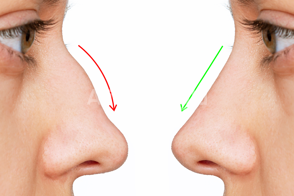
Pollybeak Deformity After Rhinoplasty: Why Does the Nose Look Bulky Above the Tip? A successful rhinoplasty is not determined solely by how straight the nasal bridge appears. The relationship between the bridge and the nasal tip also plays an important role in creating a balanced profile. When the area immediately above the tip becomes unusually prominent, the nose may develop a rounded contour that looks different from the intended surgical result. This appearance is sometimes associated with pollybeak deformity after rhinoplasty, a condition characterized by excessive fullness in the region between the nasal bridge and tip. Patients may describe their nose as looking bulky, curved, or insufficiently defined, even when the upper portion of the bridge appears relatively straight. However, a swollen supratip region does not automatically indicate a permanent rhinoplasty complication. Temporary postoperative swelling, scar tissue accumulation, residual cartilage, and changes in nasal tip support can produce similar appearances. Recognizing the anatomical differences behind these changes is essential for understanding whether the nose is still healing or whether further treatment may eventually be necessary. What Is Pollybeak Deformity and How Does It Affect the Nasal Profile? Pollybeak deformity is a postoperative nasal contour irregularity in which the supratip region appears excessively prominent in relation to the nasal tip. The supratip is the area immediately above the tip, where the lower nasal bridge transitions into the tip-defining structures. In a well-proportioned nasal profile, this transition creates a balanced relationship between the nasal dorsum and the tip. Depending on the patient’s anatomy and aesthetic preferences, a subtle change in contour may help distinguish the nasal tip from the bridge. When pollybeak deformity develops, the normal relationship between these structures becomes disrupted. Instead of a smooth transition, the lower portion of the nasal bridge may develop an outward curve. The resulting convex profile resembles the shape of a parrot’s beak, which explains the medical term pollybeak deformity. The prominence does not necessarily mean that the nasal tip itself has become larger. In many cases, the problem involves excessive supratip projection, inadequate nasal tip support, or a combination of both. For example, if the nasal tip gradually loses projection after surgery, the tissue immediately above it may appear disproportionately prominent. The supratip region can therefore look bulky even without a substantial increase in its actual volume. Alternatively, residual cartilage or excessive scar tissue may directly increase fullness above the tip, creating a similar external appearance through a different anatomical mechanism. This distinction matters because pollybeak deformity describes the visible contour rather than identifying one specific underlying cause. Patients may notice that the nasal tip appears less defined, the lower bridge looks unusually rounded, or the profile lacks the refinement they expected after rhinoplasty. In some individuals, the irregularity is most noticeable from the side. In others, changes in tip definition may also affect the appearance of the nose from an oblique angle. Pollybeak deformity can occur after both primary and revision rhinoplasty, regardless of whether an open or closed surgical approach was used. Nevertheless, early postoperative fullness should not be confused with an established deformity. The nasal tip and supratip region can retain swelling for an extended period, particularly in patients with thicker skin. A definitive assessment requires consideration of the healing timeline, nasal skin characteristics, cartilage framework, and the relationship between the nasal bridge and tip. Understanding these anatomical features provides the foundation for identifying why pollybeak deformity develops and which treatment approaches may be appropriate. Why Does Pollybeak Deformity Develop After Rhinoplasty? The development of pollybeak deformity after rhinoplasty is closely related to how the nasal cartilage, soft tissues, and supporting structures respond to surgical changes. Although the deformity produces a recognizable bulge above the nasal tip, the anatomical reasons behind this appearance can vary considerably between patients. In some cases, the supratip region becomes physically thicker. In others, the nasal tip loses projection, making the area above it appear unusually prominent. Understanding this distinction is essential because pollybeak deformity can result from either excess tissue volume or an imbalance in nasal structural support. One of the most important causes is excessive scar tissue formation in the supratip region. Following rhinoplasty, the body produces collagen and fibrous tissue as part of its natural healing response. When this process becomes pronounced, scar tissue may accumulate between the skin and the underlying nasal framework. This additional tissue can gradually create a rounded appearance along the lower nasal bridge. The condition is sometimes described as soft-tissue pollybeak deformity, particularly when fibrous tissue accumulation is the primary cause of supratip fullness. Patients with thicker nasal skin may be more susceptible to persistent fullness because their soft-tissue envelope can be less responsive to subtle changes in the underlying cartilage. However, thick nasal skin does not automatically mean that pollybeak deformity will develop. Another important mechanism involves residual cartilage along the cartilaginous nasal dorsum. During rhinoplasty, the surgeon may reshape the bridge by modifying bone and cartilage. If the cartilaginous portion remains disproportionately prominent compared with the newly established nasal profile, the supratip region may develop a visible convexity. This form of pollybeak deformity is primarily structural rather than being caused by scar tissue alone. The distinction is particularly important because excess cartilage cannot be corrected simply by waiting for postoperative swelling to disappear. Changes in nasal tip support represent another potential cause. The nasal tip maintains its position through a complex arrangement of cartilage, connective tissue, and supporting anatomical relationships. When these structures are altered during surgery, the tip may become less stable. If adequate support is not maintained, the nasal tip can gradually lose projection or rotate downward during healing. As the tip settles, the supratip region may appear increasingly prominent, even when its actual volume has not changed significantly. This explains why some patients develop a pollybeak appearance despite having undergone substantial reduction of the nasal bridge. In these situations, removing additional tissue from the supratip region without addressing the underlying support problem may fail to correct the imbalance. Over-resection or weakening of

Capsular Contracture After Breast Implants: Why Do Breasts Become Hard or Change Shape?

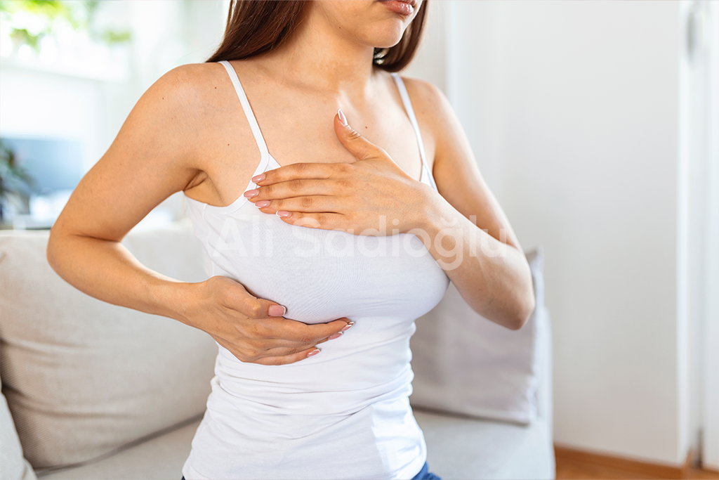
Capsular Contracture After Breast Implants: Why Do Breasts Become Hard or Change Shape? Breast augmentation with implants can enhance breast volume, improve proportions, and create a fuller appearance. However, some patients experience changes in breast firmness, shape, or position months or even years after surgery. One possible explanation is capsular contracture after breast implants, a complication involving the scar tissue that naturally forms around an implant. Although the formation of a protective tissue capsule is a normal response to breast implant placement, problems can develop when this capsule becomes excessively tight. The resulting pressure may cause the breast to feel unusually firm, appear distorted, or become uncomfortable. Understanding why breast implants become hard or change shape can help patients recognize potential complications and seek appropriate medical evaluation. What Is Capsular Contracture After Breast Implants? Capsular contracture occurs when the scar tissue surrounding a breast implant thickens, tightens, or contracts excessively, placing pressure on the implant and surrounding breast tissues. Whenever a breast implant is inserted, the body recognizes it as a foreign material and naturally develops a thin layer of fibrous tissue around it. This structure, known as the implant capsule, helps separate the implant from surrounding tissues. In most patients, the capsule remains relatively soft and flexible, allowing the breast to maintain a natural appearance and feel. However, when the capsule becomes abnormally firm or contracts around the implant, the breast may begin to feel harder or develop visible changes in shape. The severity of capsular contracture is commonly evaluated using the Baker classification system. Baker Grade I describes a breast that remains soft and appears natural, without clinically significant contracture. Baker Grade II indicates increased firmness while the breast generally maintains a normal appearance. Baker Grade III involves noticeable firmness accompanied by visible distortion of the breast. Baker Grade IV represents more severe contracture, typically associated with significant hardness, visible changes in shape, and pain. Not every patient experiences the same symptoms. Some notice only a gradual increase in firmness, while others develop discomfort or substantial changes in breast contour. Capsular contracture can affect one or both breasts, and the severity may differ between sides. The condition can develop during the early postoperative period or appear several years after breast augmentation. Why Do Breasts Become Hard or Change Shape After Implant Surgery? The development of hard breasts after breast augmentation is primarily associated with excessive tightening of the fibrous capsule surrounding the implant. Although the exact mechanisms are not completely understood, several biological and surgical factors may contribute to this process. One important factor is the body’s inflammatory response. Following implant placement, the surrounding tissues undergo a natural healing process. In some cases, persistent inflammation may stimulate increased collagen production and cause the capsule to become thicker or less flexible. Bacterial biofilm formation has also been investigated as a possible contributing factor. A biofilm is a thin community of microorganisms that can adhere to an implant surface. Even without obvious signs of infection, bacterial contamination may contribute to prolonged inflammation and increase the likelihood of abnormal capsule formation. Another potential factor is bleeding or hematoma formation after surgery. When blood accumulates around an implant, the resulting inflammatory response may increase the risk of excessive scar tissue development. Implant characteristics and surgical techniques can also influence capsular contracture risk. Factors such as implant placement, implant surface characteristics, previous surgery, radiation exposure, and individual healing responses may affect how the surrounding tissues react. However, the relationship between these factors and capsular contracture is complex, and no single factor explains every case. In patients who have undergone previous breast implant surgery, existing scar tissue and changes in the implant pocket may also contribute to recurrent contracture. As the capsule tightens, it compresses the implant and can alter its position or shape. This may cause the breast to appear unusually round, elevated, or asymmetric. A breast that gradually becomes harder, higher, or visibly distorted should be assessed by a qualified plastic surgeon, particularly when these changes are accompanied by discomfort. What Are the Symptoms of Capsular Contracture? The symptoms of capsular contracture after breast implants vary according to the severity of the condition and the degree of pressure exerted on the implant. In mild cases, patients may notice that one breast feels slightly firmer than the other. The breast may still look natural, and there may be little or no discomfort. As the capsule becomes tighter, changes in breast shape and position may become more noticeable. One common sign is progressive breast firmness. Instead of feeling relatively soft and mobile, the breast may develop an unusually rigid texture. Patients sometimes describe the implant as feeling tightly compressed beneath the skin. Another characteristic symptom is a change in breast contour. The affected breast may appear rounder, higher, or more elevated than the opposite breast. The lower portion of the breast may lose some of its natural fullness, while the upper pole becomes more prominent. Visible asymmetry may develop when capsular contracture affects only one side or progresses differently between the breasts. In more advanced cases, patients may experience tightness, tenderness, or persistent breast discomfort. Pain is not present in every case, but it becomes more common with severe contracture. Other possible signs include reduced implant mobility, a sensation of pressure, and changes in the position of the nipple relative to the breast mound. It is important to recognize that breast firmness does not always indicate capsular contracture. Temporary swelling, postoperative inflammation, implant displacement, and other breast conditions may produce similar symptoms. A clinical examination is therefore necessary to determine the underlying cause. Dr. Ali Sadioğlu evaluates changes in breast firmness, implant position, and overall breast contour to help distinguish capsular contracture from other complications following breast augmentation. When appropriate, ultrasound or magnetic resonance imaging (MRI) may be recommended to assess implant integrity, surrounding tissues, or other possible abnormalities. New or persistent swelling, a breast mass, or a significant change in breast shape should not automatically be attributed to capsular contracture. These findings