- moderator10
- 0 Comments
Pollybeak Deformity After Rhinoplasty: Why Does the Nose Look Bulky Above the Tip?
A successful rhinoplasty is not determined solely by how straight the nasal bridge appears. The relationship between the bridge and the nasal tip also plays an important role in creating a balanced profile. When the area immediately above the tip becomes unusually prominent, the nose may develop a rounded contour that looks different from the intended surgical result.
This appearance is sometimes associated with pollybeak deformity after rhinoplasty, a condition characterized by excessive fullness in the region between the nasal bridge and tip. Patients may describe their nose as looking bulky, curved, or insufficiently defined, even when the upper portion of the bridge appears relatively straight.
However, a swollen supratip region does not automatically indicate a permanent rhinoplasty complication. Temporary postoperative swelling, scar tissue accumulation, residual cartilage, and changes in nasal tip support can produce similar appearances.
Recognizing the anatomical differences behind these changes is essential for understanding whether the nose is still healing or whether further treatment may eventually be necessary.

What Is Pollybeak Deformity and How Does It Affect the Nasal Profile?
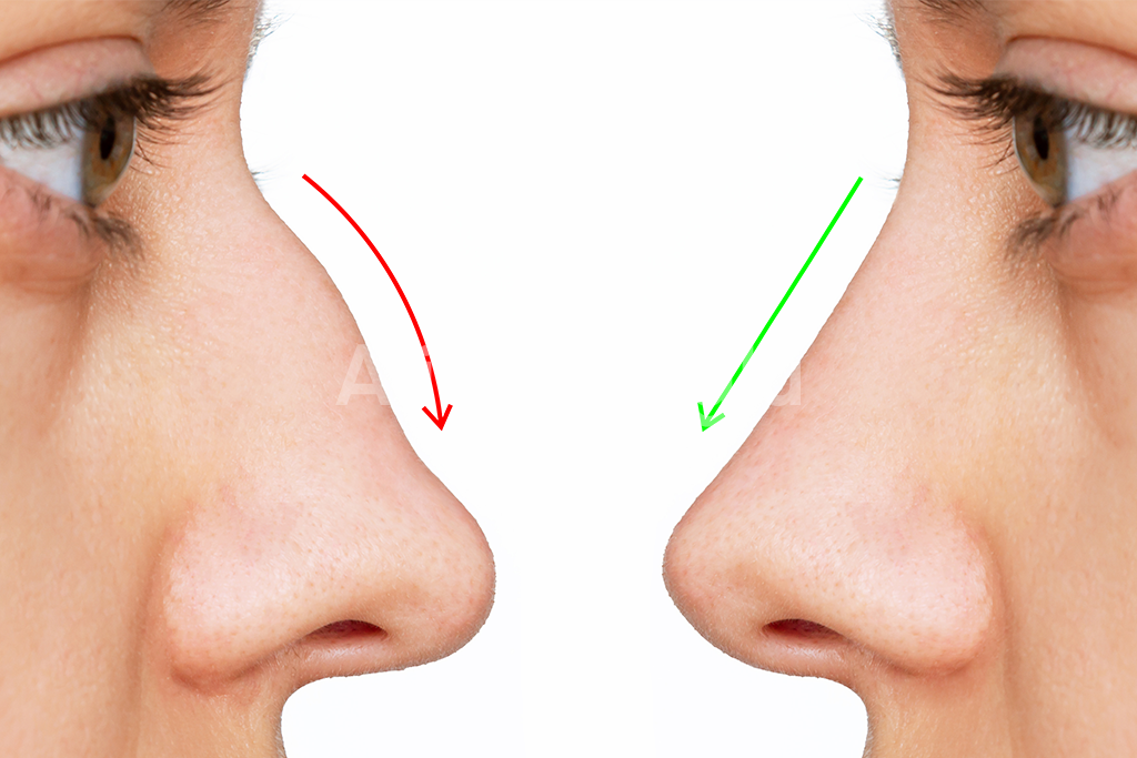
Pollybeak deformity is a postoperative nasal contour irregularity in which the supratip region appears excessively prominent in relation to the nasal tip. The supratip is the area immediately above the tip, where the lower nasal bridge transitions into the tip-defining structures.
In a well-proportioned nasal profile, this transition creates a balanced relationship between the nasal dorsum and the tip. Depending on the patient’s anatomy and aesthetic preferences, a subtle change in contour may help distinguish the nasal tip from the bridge.
When pollybeak deformity develops, the normal relationship between these structures becomes disrupted. Instead of a smooth transition, the lower portion of the nasal bridge may develop an outward curve.
The resulting convex profile resembles the shape of a parrot’s beak, which explains the medical term pollybeak deformity.
The prominence does not necessarily mean that the nasal tip itself has become larger. In many cases, the problem involves excessive supratip projection, inadequate nasal tip support, or a combination of both.
For example, if the nasal tip gradually loses projection after surgery, the tissue immediately above it may appear disproportionately prominent. The supratip region can therefore look bulky even without a substantial increase in its actual volume.
Alternatively, residual cartilage or excessive scar tissue may directly increase fullness above the tip, creating a similar external appearance through a different anatomical mechanism.
This distinction matters because pollybeak deformity describes the visible contour rather than identifying one specific underlying cause.
Patients may notice that the nasal tip appears less defined, the lower bridge looks unusually rounded, or the profile lacks the refinement they expected after rhinoplasty.
In some individuals, the irregularity is most noticeable from the side. In others, changes in tip definition may also affect the appearance of the nose from an oblique angle.
Pollybeak deformity can occur after both primary and revision rhinoplasty, regardless of whether an open or closed surgical approach was used.
Nevertheless, early postoperative fullness should not be confused with an established deformity. The nasal tip and supratip region can retain swelling for an extended period, particularly in patients with thicker skin.
A definitive assessment requires consideration of the healing timeline, nasal skin characteristics, cartilage framework, and the relationship between the nasal bridge and tip.
Understanding these anatomical features provides the foundation for identifying why pollybeak deformity develops and which treatment approaches may be appropriate.
Why Does Pollybeak Deformity Develop After Rhinoplasty?
The development of pollybeak deformity after rhinoplasty is closely related to how the nasal cartilage, soft tissues, and supporting structures respond to surgical changes. Although the deformity produces a recognizable bulge above the nasal tip, the anatomical reasons behind this appearance can vary considerably between patients.
In some cases, the supratip region becomes physically thicker. In others, the nasal tip loses projection, making the area above it appear unusually prominent. Understanding this distinction is essential because pollybeak deformity can result from either excess tissue volume or an imbalance in nasal structural support.
One of the most important causes is excessive scar tissue formation in the supratip region. Following rhinoplasty, the body produces collagen and fibrous tissue as part of its natural healing response. When this process becomes pronounced, scar tissue may accumulate between the skin and the underlying nasal framework.
This additional tissue can gradually create a rounded appearance along the lower nasal bridge. The condition is sometimes described as soft-tissue pollybeak deformity, particularly when fibrous tissue accumulation is the primary cause of supratip fullness.
Patients with thicker nasal skin may be more susceptible to persistent fullness because their soft-tissue envelope can be less responsive to subtle changes in the underlying cartilage. However, thick nasal skin does not automatically mean that pollybeak deformity will develop.
Another important mechanism involves residual cartilage along the cartilaginous nasal dorsum. During rhinoplasty, the surgeon may reshape the bridge by modifying bone and cartilage. If the cartilaginous portion remains disproportionately prominent compared with the newly established nasal profile, the supratip region may develop a visible convexity.
This form of pollybeak deformity is primarily structural rather than being caused by scar tissue alone. The distinction is particularly important because excess cartilage cannot be corrected simply by waiting for postoperative swelling to disappear.
Changes in nasal tip support represent another potential cause.
The nasal tip maintains its position through a complex arrangement of cartilage, connective tissue, and supporting anatomical relationships. When these structures are altered during surgery, the tip may become less stable.
If adequate support is not maintained, the nasal tip can gradually lose projection or rotate downward during healing. As the tip settles, the supratip region may appear increasingly prominent, even when its actual volume has not changed significantly.
This explains why some patients develop a pollybeak appearance despite having undergone substantial reduction of the nasal bridge.
In these situations, removing additional tissue from the supratip region without addressing the underlying support problem may fail to correct the imbalance.
Over-resection or weakening of important tip-support structures can therefore contribute to an unfavorable relationship between the nasal dorsum and nasal tip.
Postoperative healing also influences the final nasal contour. Swelling, tissue contraction, and scar maturation do not always occur uniformly across the nose.
The supratip region may retain fluid longer than neighboring areas, temporarily creating a rounded profile. Although this appearance can resemble pollybeak deformity, persistent swelling and permanent structural deformity are not the same condition.
Previous nasal surgery may further complicate the healing process. Patients undergoing revision rhinoplasty often have altered cartilage anatomy, existing scar tissue, and reduced flexibility in the surrounding soft tissues.
These changes can make the relationship between the nasal framework and skin envelope more difficult to predict.
In certain cases, multiple anatomical factors contribute to pollybeak deformity simultaneously. A patient may have residual dorsal cartilage, moderate scar tissue accumulation, and reduced nasal tip projection, all influencing the final appearance.
For this reason, determining the cause requires more than examining the visible bulge.
A detailed assessment should consider supratip tissue thickness, cartilage prominence, nasal tip projection, tip rotation, skin characteristics, and postoperative healing progression.
Identifying which structures are responsible for the contour irregularity is the foundation of selecting an appropriate correction strategy while preserving both nasal appearance and breathing function.
How Can You Distinguish Pollybeak Deformity from Normal Postoperative Swelling?
Noticing a rounded or unusually prominent area above the nasal tip after rhinoplasty can be concerning, especially when the nose does not immediately resemble the expected surgical result. However, supratip swelling after rhinoplasty and true pollybeak deformity can look remarkably similar, particularly during the early stages of recovery.
The key difference lies in what causes the fullness and how the nasal contour changes as healing progresses. While postoperative swelling is a temporary response to surgical trauma, an established pollybeak deformity may involve excessive scar tissue, residual cartilage, or inadequate nasal tip projection.
Following rhinoplasty, the tissues surrounding the nasal framework undergo inflammation, fluid accumulation, and gradual remodeling. These processes are necessary for healing but can temporarily obscure the definition created during surgery.
The nasal tip and supratip region are particularly susceptible to prolonged swelling because of their soft-tissue characteristics and the extent of surgical manipulation.
During the first few weeks after rhinoplasty, a bulky appearance above the nasal tip is often associated with normal postoperative edema. The lower portion of the nose may look rounded, and the transition between the bridge and tip may appear less distinct than expected.
As recovery continues, swelling generally decreases, although the improvement is not always uniform. Some areas may become more defined earlier, while the supratip region remains relatively full.
Patients may also notice that nasal swelling fluctuates throughout the day. Temporary changes in fluid retention can make the nose appear more swollen at certain times, particularly during the initial healing period.
These fluctuations do not necessarily indicate that the surgical result is deteriorating.
By contrast, structural pollybeak deformity may produce a persistent convexity that does not substantially improve as postoperative swelling resolves.
When excess cartilage is responsible, the prominence reflects the underlying nasal framework rather than temporary fluid accumulation. Similarly, insufficient tip support can create a lasting imbalance between the nasal dorsum and tip.
Scar-related pollybeak deformity may be more difficult to distinguish from swelling because both involve changes in the soft tissues.
As healing progresses, excessive fibrous tissue can contribute to persistent supratip fullness. Unlike ordinary edema, this tissue may not disappear simply through the natural reduction of postoperative swelling.
However, the appearance or firmness of the supratip region alone cannot reliably establish whether the problem is swelling, scar tissue, or cartilage prominence.
The timing of the assessment is therefore particularly important.
During the first three months, the nose may undergo noticeable changes as inflammation decreases and the surrounding tissues begin adapting to their new position.
Between three and six months, many patients experience further improvement in nasal definition. Nevertheless, residual swelling may remain visible, especially around the tip.
The final nasal contour commonly requires approximately 12 months to mature, and some patients may continue experiencing refinement beyond this period.
Individuals with thicker skin or those undergoing revision rhinoplasty may require additional healing time before the result can be assessed reliably.
This does not mean that every patient must wait a full year before discussing a concern. Progressive deformity, unexpected changes, breathing difficulties, or other unusual symptoms should be evaluated when they arise.
A plastic surgeon can examine the relationship between the nasal bridge and tip, assess tissue thickness, and review changes documented throughout recovery.
Comparing standardized postoperative photographs taken at different stages can help determine whether supratip fullness is gradually improving or remaining relatively unchanged.
Clinical examination may also reveal whether the nasal tip has adequate structural support or whether the contour suggests residual cartilage prominence.
In selected cases, additional assessment may be necessary to clarify the underlying anatomy.
Patients should avoid attempting to correct persistent fullness through aggressive massage, pressure, or unprescribed treatments, as these measures may interfere with healing or cause unnecessary tissue irritation.
The most reliable way to distinguish postoperative swelling from pollybeak deformity is to evaluate the nasal contour over time while considering the underlying cartilage structure, soft-tissue response, and tip support.
Recognizing these differences helps prevent unnecessary early intervention while allowing genuine structural problems to be identified and managed appropriately.
Can Pollybeak Deformity Be Corrected Without Revision Rhinoplasty?
Not every patient who develops fullness above the nasal tip requires another operation. In certain circumstances, pollybeak deformity after rhinoplasty may improve without revision surgery, particularly when the prominent supratip contour is associated with postoperative swelling or excessive soft-tissue scarring rather than an underlying cartilage abnormality.
The possibility of non-surgical correction depends primarily on identifying what is responsible for the unusual nasal profile. A rounded supratip caused by temporary inflammation requires a different approach from a deformity involving residual cartilage or inadequate nasal tip support.
Non-surgical treatment is most likely to be helpful when the problem originates in the soft tissues rather than the structural framework of the nose.
During the early healing period, postoperative swelling can create the impression that the area above the nasal tip is excessively prominent. As inflammation gradually decreases, the relationship between the nasal bridge and tip may become more defined without additional intervention.
For this reason, careful observation is often the most appropriate initial approach when supratip fullness appears during the first months after rhinoplasty.
The healing process should be evaluated through scheduled examinations rather than relying on the appearance of the nose at a single point in time. Changes in swelling, skin thickness, and tissue firmness can provide useful information about whether the contour is continuing to improve.
However, waiting is not necessarily the best approach in every situation. A progressively worsening deformity, significant breathing difficulty, or an unexpected change in nasal structure warrants timely assessment.
When excessive scar tissue contributes to the fullness, corticosteroid injections may be considered as a non-surgical treatment for selected cases of soft-tissue pollybeak deformity.
These injections are intended to reduce localized inflammatory and fibrotic tissue responses. By decreasing excessive soft-tissue thickening, they may help improve the definition of the supratip region.
The effectiveness of steroid treatment depends on the characteristics of the tissue, the timing of intervention, and the underlying cause of the deformity.
Steroid injections cannot remove residual cartilage, restore weakened nasal tip support, or reliably correct a structural pollybeak deformity.
They also require careful administration. Excessive dosage or inappropriate placement can contribute to skin thinning, changes in pigmentation, tissue atrophy, or visible contour depressions.
For these reasons, corticosteroid treatment should only be performed when a qualified surgeon determines that the potential benefits outweigh the risks.
Another approach sometimes used during postoperative recovery involves nasal taping.
In selected patients, a surgeon may recommend carefully applied tape to help manage postoperative swelling and support the soft-tissue envelope during healing.
However, the effectiveness of taping varies, and nasal taping should not be considered a definitive treatment for established pollybeak deformity.
It cannot reshape prominent cartilage or compensate for insufficient structural support.
Patients should also avoid applying excessive pressure, performing aggressive nasal massage, or attempting home treatments without medical guidance. Such measures may irritate healing tissues and are unlikely to correct the anatomical causes of persistent supratip fullness.
A particularly important distinction involves structural versus soft-tissue pollybeak deformity.
When the supratip prominence results from excess cartilage along the lower nasal dorsum, non-surgical measures generally cannot eliminate the underlying projection.
Similarly, when the nasal tip has lost support and settled into a lower position, reducing swelling alone will not restore the original relationship between the bridge and tip.
In these circumstances, revision rhinoplasty may be necessary to correct the structural imbalance.
The decision to pursue revision surgery should generally be made after sufficient healing has occurred, unless a specific medical concern requires earlier intervention.
For most elective aesthetic revisions, surgeons commonly recommend allowing approximately 12 months after the original rhinoplasty before proceeding, although the appropriate timing varies with the patient’s anatomy and healing progress.
Dr. Ali Sadioğlu assesses supratip tissue characteristics, nasal tip projection, cartilage support, and postoperative changes when determining whether non-surgical management may be appropriate or whether structural correction should be considered.
The goal is to avoid unnecessary surgery when the appearance may still improve naturally, while recognizing situations in which continued observation is unlikely to resolve the deformity.
Non-surgical pollybeak correction has clear limitations, and the most appropriate treatment depends on whether the visible fullness is caused by swelling, scar tissue, residual cartilage, or loss of nasal tip support.